Research Interests
Electron cryo-microscopy (cryo-EM) is a versatile technique to visualize the three-dimensional (3D) structure of macromolecules and their assemblies. With cryo-EM, it is now possible to study the atomic structure of assemblies with molecular masses as low as ~100 kDa (for example, a membrane protein) or as high as hundreds of MDa (for example, a large virus). The assemblies can be visualized single-fold, thus avoiding the need for crystals. This "single-particle" approach requires extremely small amounts of material, typically only a few tens of picomoles.
Our laboratory develops new methods for cryo-EM to achieve the highest possible resolution (2 Å or better). We have established a new movie imaging protocol [1] that makes use of a new type of camera, the direct electron detector. Using this detector, the total electron exposure used for an image can be distributed over many short movie frames rather than a single long exposure. Any sample movement that occurs during the exposure (for example, beam-induced motion) can subsequently be tracked in the movies and “undone” by aligning the movie frames to each other [2], thereby producing a final image that is free of blurring.
More recently, we started developing a method to detect molecules and assemblies in situ, inside cells (collaborator: Denk lab). A full understanding of molecular mechanisms in biology requires the cellular context to provide a complete spatial understanding of local environments and preserve weak and transient interactions otherwise missed. Electron cryo-tomography was developed to visualize molecules in situ and yields 3D reconstructions with features that can be identified using template matching. This has been particularly successful for large cytoplasmic complexes, such as ribosomes and proteasomes, that can be readily identified by their distinctive shapes. In more densely packed environments, however, such as nuclei or bacterial cells, molecular shapes are obscured due to proximity or direct contact with other molecules, making shape-based template matching unreliable. To address this problem, we have begun developing high-resolution template matching (HRTM) [3], which matches internal molecular features visible at high resolution. The internal features remain distinct even in crowded environments, and HRTM can therefore determine molecular identities. Our approach uses images of untilted specimens, which best preserve high-resolution signal, relying on closely matching templates to locate molecules with high accuracy.
To make our tools easily accessible to users, we have designed open-source software called cisTEM (computational imaging system for transmission electron microscopy) to process cryo-EM data [4]. cisTEM features a graphical user interface for submitting jobs, monitoring progress, and displaying results. It implements a full processing pipeline including all steps required for single-particle cryo-EM, including movie processing based on Unblur, CTF determination based on CTFFind4, maximum-likelihood 2D classification, ab-initio 3D reconstruction, auto-refinement, 3D classification and map sharpening. cisTEM is optimized for CPU workstations and allows fast and efficient processing without the need for large computer clusters or specialized GPU hardware.
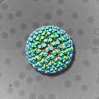
We apply the single-particle technique to assemblies that are difficult to study by more traditional techniques such as X-ray crystallography and nuclear magnetic resonance (NMR). For example, membrane proteins are usually too large for NMR analysis or are difficult to crystallize for X-ray crystallography. Large protein assemblies pose additional problems because they can undergo constant changes in composition and conformation. An example is the ribosome (collaborator: Korostelev lab), which undergoes conformational changes during translation and binds to different factors and ligands. Another class of protein assemblies that do not usually form crystals includes fibers and filaments, such as amyloid fibrils (collaborator: Fändrich lab). We also study highly symmetrical viruses that represent ideal test specimens for the development of new image processing techniques.


